Quantifying Key Component Proteins within Kinetochore Complex in Living Cells Achieved by Researchers
A major breakthrough has been achieved by researchers at the University of Osaka who have successfully quantified the number of key component proteins within the kinetochore complex in...
A major breakthrough has been achieved by researchers at the University of Osaka who have successfully quantified the number of key component proteins within the kinetochore complex in living cells. This achievement has been published in the Journal of Cell Biology and is set to revolutionize our understanding of how these proteins work together.
The kinetochore, a protein structure critical for life and growth, enables the accurate division of genetic material into two daughter cells. However, challenges have long hindered researchers from grasping the molecular organization of this complex within living cells. To rectify this, an improved fluorescence-based method has been developed to count these proteins in real-time.
As a cell prepares to divide into two, its genetic material is duplicated and paired at the centromere region. The kinetochore assembles at this precise location and serves as the attachment point for chromosomes to link with the spindle structure running throughout the cell. This intricate process ensures that each new cell receives an accurate copy of the genetic material.
The failure of chromosomes to attach accurately to the spindle can result in errors linked to various diseases, making the accurate division of genetic material a critical aspect of life and growth. To address this issue, researchers have been working tirelessly to develop methods for measuring the molecular organization of the kinetochore within living cells.
In mitotic chicken DT40 cells, researchers employed fluorescence correlation spectroscopy-calibrated molecular counting to study the kinetochore complex.
The kinetochore consists of two main protein networks: the CCAN forming the inner kinetochore and the KMN forming the outer kinetochore. Among the CCAN proteins measured, CENP-C was found to be present at a significantly higher copy number than the others. This discrepancy in copy numbers highlights the complexity of the kinetochore's molecular organization.
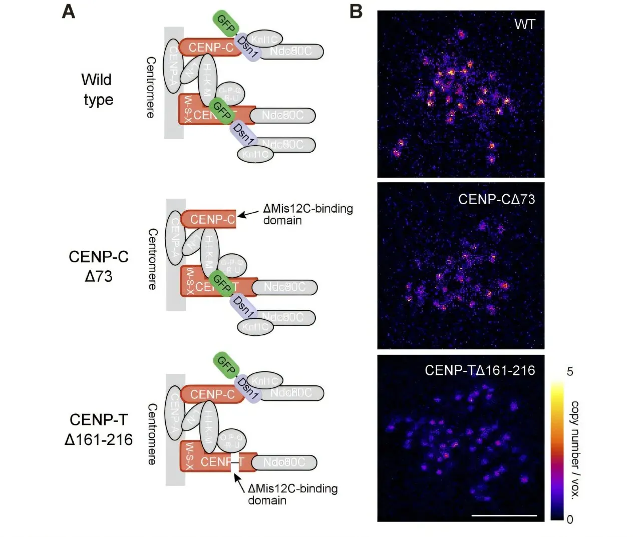
The attachment mechanism between the inner and outer kinetochore complexes involves two redundant binding sites on CENP-C and another CCAN protein, CENP-T. Both these proteins have specific binding sites for the Mis12C protein complex. The presence of these binding sites ensures a secure attachment between the two complexes.
Researchers found that removing one of the Mis12C binding sites from either CENP-C or CENP-T does not impede the cell's ability to divide successfully. In fact, sufficient Mis12C can still bind to the other CENP protein, allowing for the recruitment of KMN components and maintaining the integrity of the division process.
The research team found that a minimum of 15 molecules of Mis12C and over 30 molecules of Ndc80C are necessary for accurate cell division to occur.
According to senior author Tatsuo Fukagawa, the high number of Ndc80C molecules provides a stable platform for attachment to microtubules, with an estimated 8-10 molecules per microtubule. This suggests that sufficient quantities of these proteins are crucial for maintaining the integrity of the cell division process.
The study sheds new light on the complex process of creating structures essential for accurate cell division, a fundamental aspect of cellular growth and development.
Facts based on reporting originally published by Phys.org.
You may republish this story, in full or in part, if you credit News Central Site and link to it (licence CC BY 4.0). Photos are not included.