Plant Proteomics Advances with New Method Developed by University Researchers
A team of scientists at the University of Delaware has developed a method to study proteins inside intact plant cells without extensive chemical processing.
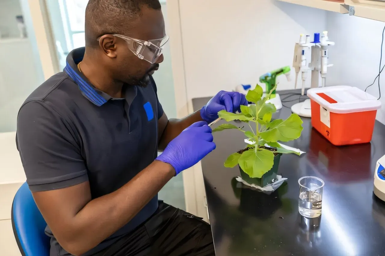
Scientists have made significant strides in understanding human biology through proteomics, the study of proteins within cells. However, similar advances in plant biology have been hindered by a crucial barrier: protecting plant tissues from unwanted substances while keeping essential components intact.
Plant cells are encased in hard exterior shells and thick cell walls that shield their internal workings from external factors. This protective mechanism is vital for plants but also presents a significant challenge for researchers seeking to study plant proteins using proteomics.
Researchers have been working to overcome this hurdle, which has hindered the field of plant proteomics. The complex structure of plant cells makes it difficult to access and analyze the proteins within.
A team of University of Delaware researchers has developed a new method that allows them to digest proteins inside intact plant cells. This breakthrough has the potential to revolutionize our understanding of plant biology and disease mechanisms.
The validated method, published in the journal Plant Physiology, promises to open up new avenues for studying plant health and disease. The research is expected to have far-reaching implications for agriculture and beyond.
The new method developed by researchers allows for the study of proteins involved in plant health and disease without the need for extensive chemical processing.
Traditionally, extracting proteins from plant tissue has been a labor-intensive process that can take days to complete, with valuable material often lost along the way. This involves grinding up the tissue using a mortar and pestle before putting it through a series of chemical steps to isolate the desired proteins.
In contrast, Yanbao Yu's approach treats intact plant tissue with methanol, which makes it possible to study proteins within cells while preserving crucial information. This method acts as a sort of "blast freezer that freezes the cellular biology in place for further examination.
By using this new approach, researchers can identify biomarkers that indicate specific bacterial or fungal infections and potentially develop treatments to help plant cells recover from disease. This could have significant implications for agriculture, where infection is a major economic concern.
The method's ability to preserve valuable material and provide detailed insights into cellular biology makes it an attractive alternative to traditional extraction methods.
The new method of methanol fixation allows researchers to break open cells without altering the original cellular pathology, making it possible to examine the exact proteins inside.
This approach is a departure from traditional methods used in proteomics analysis, where cells are typically broken apart to work directly with proteins. However, Yu's team has found that preserving the cell structure can be beneficial for certain types of research.
By using methanol fixation, the team was able to analyze the proteins within intact cells, rather than destroying them first. This approach allows researchers to use a chemical knife" to cut into the cells and examine their contents without causing damage.
The sensitivity and robustness of this method enable researchers to accurately identify large numbers of proteins from even small samples.
The researchers were interested in understanding what makes a fungus pathogenic, as not all fungi cause disease in plants. To address this question, they studied how plant protein profiles change when exposed to both disease-causing and harmless fungi.
This study revealed distinct differences in the proteins present in plants infected with disease-causing versus harmless fungi. The findings suggest that certain proteins are specific markers of fungal pathogenicity.
Plant extracellular vesicles, which facilitate cell-to-cell communication, were found to be enriched with immune-related proteins. This implies an active role for these vesicles in plant defense against pathogens.
In contrast, the researchers discovered that fungal pathogens release extracellular vesicles containing poisonous proteins that can hijack cellular processes and trigger infection. The data also showed that plants respond differently to disease-causing and harmless fungi at a molecular level.
The new method has been successfully applied not only to plants but also to various other biological systems, including model organisms such as Caenorhabditis elegans worms, frog embryos, sea urchins, bacterial cells, neuroblastoma cells, and yeasts.
The new method developed by Yu at the University of Delaware has proven to be a valuable tool for researchers across various disciplines, with over 15 laboratories on campus utilizing it to explore different areas of study.
These studies have spanned multiple fields, including developmental biology, infection and immunology, chronic nerve pain, and more. The technology platform is licensed by CDS Analytical LLC, which supplies proteomics sample preparation consumables and instruments.
The method's potential impact has been acknowledged by Yu, who believes it will be a game changer for researchers due to its practical yet powerful approach to protein sample preparation with minimal cell quantities, including potentially single cells.
Facts based on reporting originally published by Phys.org.
You may republish this story, in full or in part, if you credit News Central Site and link to it (licence CC BY 4.0). Photos are not included.