Groundbreaking Discovery Made in Plant Biology and Disease Response Research
Scientists have developed a new method for creating glowing biological reporters that can track gene activity in living plants over extended periods.
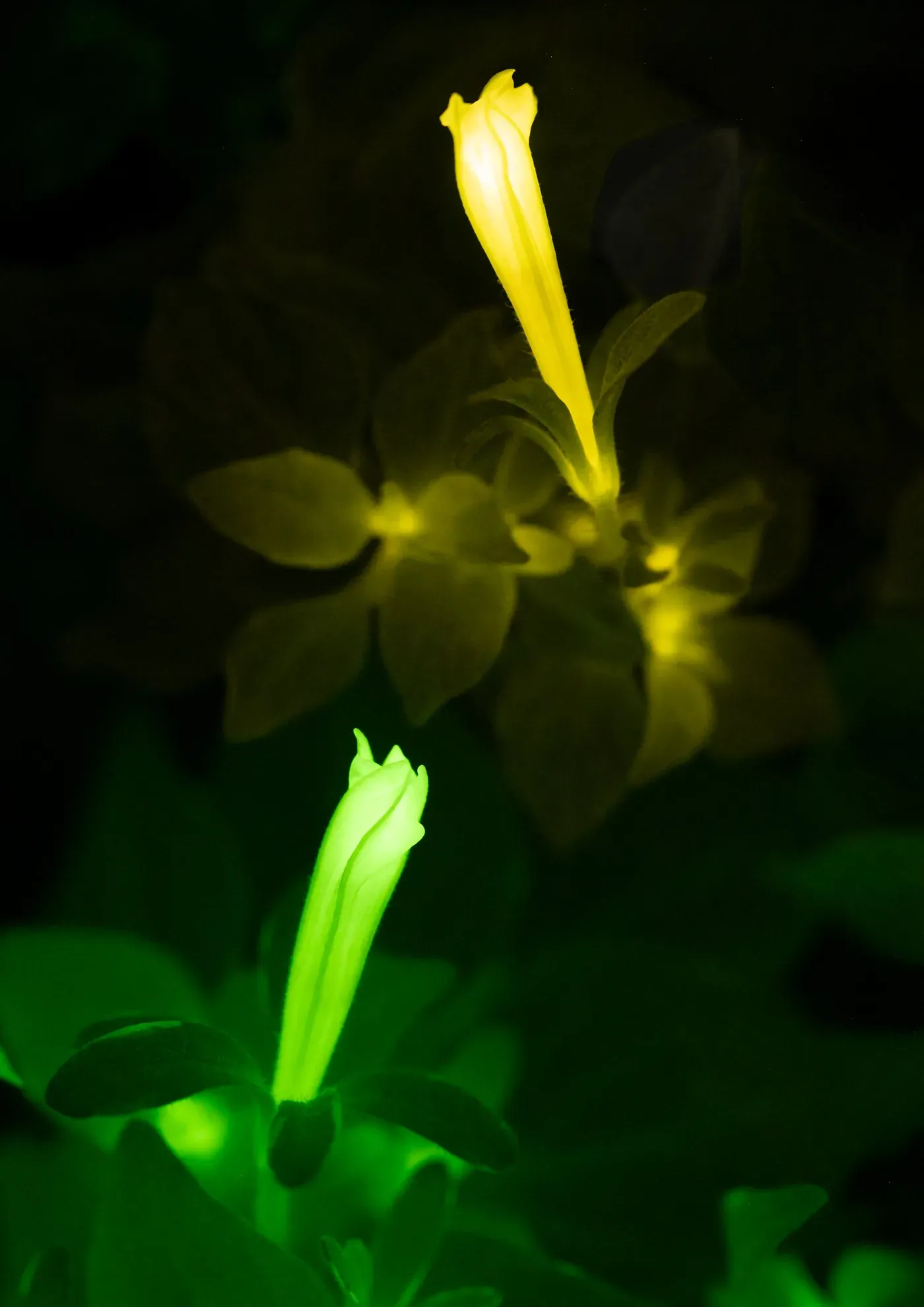
Researchers at a leading medical laboratory have made a groundbreaking discovery that could revolutionize our understanding of plant biology and disease response. By developing a new method for creating glowing biological reporters, scientists can now track gene activity in living plants over extended periods of time.
The breakthrough has the potential to transform the field of autoluminescence imaging, which allows organisms to produce their own light without the need for external chemicals or specialized equipment. The innovative approach, led by researchers working under Karen Sarkisyan's guidance, could pave the way for similar applications in animal and human disease models.
A major challenge in using glowing biological reporters has been distinguishing between changes in gene activity and shifts in an organism's physiological state. Light output can be influenced by various factors, including metabolism, temperature, oxygen levels, and more. This makes it difficult to determine whether observed changes in brightness accurately reflect biological activity or are simply a result of environmental fluctuations.
The new method developed by the researchers aims to address this issue by providing a more accurate way to track gene activity over time. By doing so, scientists can gain valuable insights into how plants respond to disease, pests, and environmental stress. This knowledge could have significant implications for fields such as agriculture, medicine, and conservation biology.
The study's findings were published in Nature Plants today, marking an important milestone in the development of autoluminescence imaging technology. The research team collaborated with international partners from Light Bio and other institutions to bring this innovative approach to life.
The researchers tackled this challenge by creating a new version of fungal luciferase that emits light in the red spectrum, which they paired with a green luminescent reporter. This dual-color approach allowed them to measure the ratio between the two colors instead of relying solely on overall brightness.
This innovative system transformed autoluminescence from a qualitative indicator into a more precise quantitative tool, providing a clearer picture of what is happening inside living organisms. By using this method, researchers can now monitor key defense and immune signaling pathways in real-time.
The team successfully demonstrated the effectiveness of this technique by tracking hormone-driven responses over extended periods in plants. They were also able to detect localized immune activity around sites of infection, even when overall brightness was affected by variations in metabolism.
The dual-color reporters showed remarkable adaptability, working seamlessly across both fungal and bacterial bioluminescence pathways. This versatility suggests that the system can be adapted for a wide range of biological applications, offering new possibilities for research and discovery.
This breakthrough technology has transformed autoluminescence into a precise tool for monitoring molecular processes in real-time. Researchers can now capture detailed information about biological activities simply by taking photographs of plants in the dark, eliminating the need for expensive equipment or invasive treatments.
The ability to continuously monitor plant health and nutrient levels could lead to the development of "sentinel" plants that provide quantitative data on environmental conditions and disease through changes in their light emission. This technology also has the potential to revolutionize the study of plant-pathogen interactions by allowing researchers to observe crop health under more realistic field conditions.
The team behind this innovation believes its underlying principles can be applied beyond plant biology, with far-reaching implications for various fields of research. They are now exploring ways to adapt similar approaches for use in animal tissues and disease models, which could enable the monitoring of hormone activity, gene regulation, and disease-related processes over extended periods using simple imaging methods.
This versatility suggests that the system can be adapted for a wide range of biological applications, offering new possibilities for research and discovery. The researchers' goal is to develop non-invasive methods for studying complex biological systems, allowing scientists to gain deeper insights into the underlying mechanisms driving these processes.
The significance of this breakthrough has not gone unnoticed by the scientific community, with senior editor Guillaume Tena highlighting its potential in an accompanying Research Briefing. He noted that the new red-shifted luciferase provides a crucial internal control, enabling researchers to distinguish genuine biological signals from metabolic fluctuations and unlocking the full potential of fungal bioluminescence for quantitative applications.
The new red-shifted luciferase has been engineered to emit a distinct color when activated by oxygen, allowing researchers to monitor metabolic activity in real-time. This bioluminescent marker is particularly useful for tracking immune responses in plants, as it can be introduced into specific cells or tissues without disrupting their function.
This innovation enables scientists to visualize the complex interactions between plants and pathogens with unprecedented clarity, shedding light on the intricate mechanisms of plant immunity. By harnessing this technology, researchers aim to develop more targeted and effective strategies for protecting crops from disease, ultimately improving food security worldwide.
Facts based on reporting originally published by Phys.org.
You may republish this story, in full or in part, if you credit News Central Site and link to it (licence CC BY 4.0). Photos are not included.