New Microscope Offers Wide Range of Imaging Tools in One System
Researchers at UT Southwestern Medical Center have developed the multi-immersion oblique plane microscope (miOPM), a light-sheet microscope that captures three-dimensional views of biological samples.
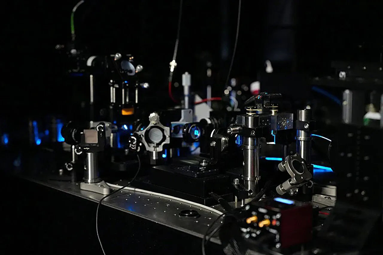
Researchers at UT Southwestern Medical Center have made significant strides in developing a new type of microscope that offers a wide range of imaging tools in one system. This innovative device is capable of capturing three-dimensional views of biological samples, providing valuable insights into the inner workings of living cells and larger tissues.
The light-sheet microscope, known as the multi-immersion oblique plane microscope (miOPM), is designed to make detailed imaging faster and simpler than ever before. According to its developers, this approach has the potential to help laboratories stretch their equipment budgets and expand their research capabilities.
Dr. Reto Fiolka, an associate professor at UT Southwestern, emphasized the versatility of the new microscope technique. Our work makes light-sheet fluorescence microscopy more user-friendly, he explained. This breakthrough could have far-reaching implications for researchers working in various fields.
The miOPM addresses a long-standing challenge in microscopy: many advanced microscopes are designed to perform only specific tasks. Studying different samples often requires separate instruments, specialized preparation, and considerable technical expertise. The new platform aims to provide a practical solution by making sophisticated 3D imaging more accessible.
By offering a single instrument that can handle multiple tasks, the miOPM has the potential to save researchers time and resources. This could be particularly beneficial for labs with limited budgets or those working on complex projects that require frequent reconfiguration of their equipment.
The development of the miOPM has been a collaborative effort between researchers from various institutions around the world. In addition to the University of Texas Southwestern Medical Center (UTSW), where the study was led by Fiolka, teams from Australia, Germany, Poland, and the UK were also involved.
Traditional light-sheet microscopes are often designed with a single purpose in mind, which can limit their versatility. For example, some models require oil immersion objectives for zooming in on tiny cellular structures, while others need to be used with water or specialized fluids to examine larger tissues or organoids. Furthermore, some microscopes must be able to scan multiple sample dishes without touching them, necessitating the use of air between the lens and sample.
The miOPM has been designed to overcome these limitations by allowing researchers to switch between different objective lenses on a single microscope. This means that labs can adapt their equipment for various experiments without having to purchase separate microscopes for each task. An adjustable zoom lens is also incorporated into the system, which helps preserve image quality when the liquid surrounding a sample is changed.
The miOPM's ability to handle different liquids and adjust its optics accordingly makes it an attractive option for researchers working on diverse projects. By being able to switch between oil, water, or air as needed, scientists can use one system for a wide range of studies without having to rebuild the internal optics of their microscope.
Like other light-sheet microscopes, the miOPM illuminates only one thin section of a sample at a time, which helps build a 3D picture while minimizing unnecessary light exposure. This approach supports rapid imaging and protects living samples from damage caused by excessive light, allowing scientists to observe biological activity as it unfolds in real-time.
The new 3D microscope has been put to the test in various applications, showcasing its versatility and efficiency.
Researchers have successfully used the platform to observe moving structures on immune cells, track activity within cells' energy-producing structures, and examine lung and liver cancer tissue. This demonstrates the system's ability to handle a range of samples and provide detailed insights into biological processes.
The microscope has also been shown to work seamlessly with standard laboratory plates containing hundreds of sample wells. This capability allows for the efficient study of multiple samples simultaneously, making it an ideal tool for researchers who need to analyze large numbers of specimens.
This flexibility could have significant implications for disease research, enabling scientists to gain a deeper understanding of how cells function and how diseases affect tissue over time. By uncovering these insights, researchers may be able to develop better diagnostic approaches and therapies.
The new technology's potential impact on patient health is also substantial, as it enables pharmaceutical researchers to test thousands of drug candidates much faster to see how they affect disease targets in 3D. This could lead to more targeted and effective treatments for a range of conditions, including Parkinson's, cancer, and respiratory diseases.
Researchers at UT Southwestern and Calico Life Sciences have successfully used two miOPM systems to capture high-resolution images of cells and tissues in various conditions. This technology has the potential to revolutionize our understanding of cellular behavior and disease mechanisms.
The imaging capabilities of these systems can be reconfigured to suit different research needs, allowing scientists to study a range of biological processes with greater precision and accuracy. As a result, this could lead to more targeted and effective treatments for various conditions, including Parkinson's, cancer, and respiratory diseases.
Facts based on reporting originally published by Phys.org.
You may republish this story, in full or in part, if you credit News Central Site and link to it (licence CC BY 4.0). Photos are not included.