Visualizing Enzyme Crucial for Cancer Drug Production
A team of researchers from Japan has successfully captured the image of SacC, an enzyme that assembles the five-ring core structure shared with trabectedin.
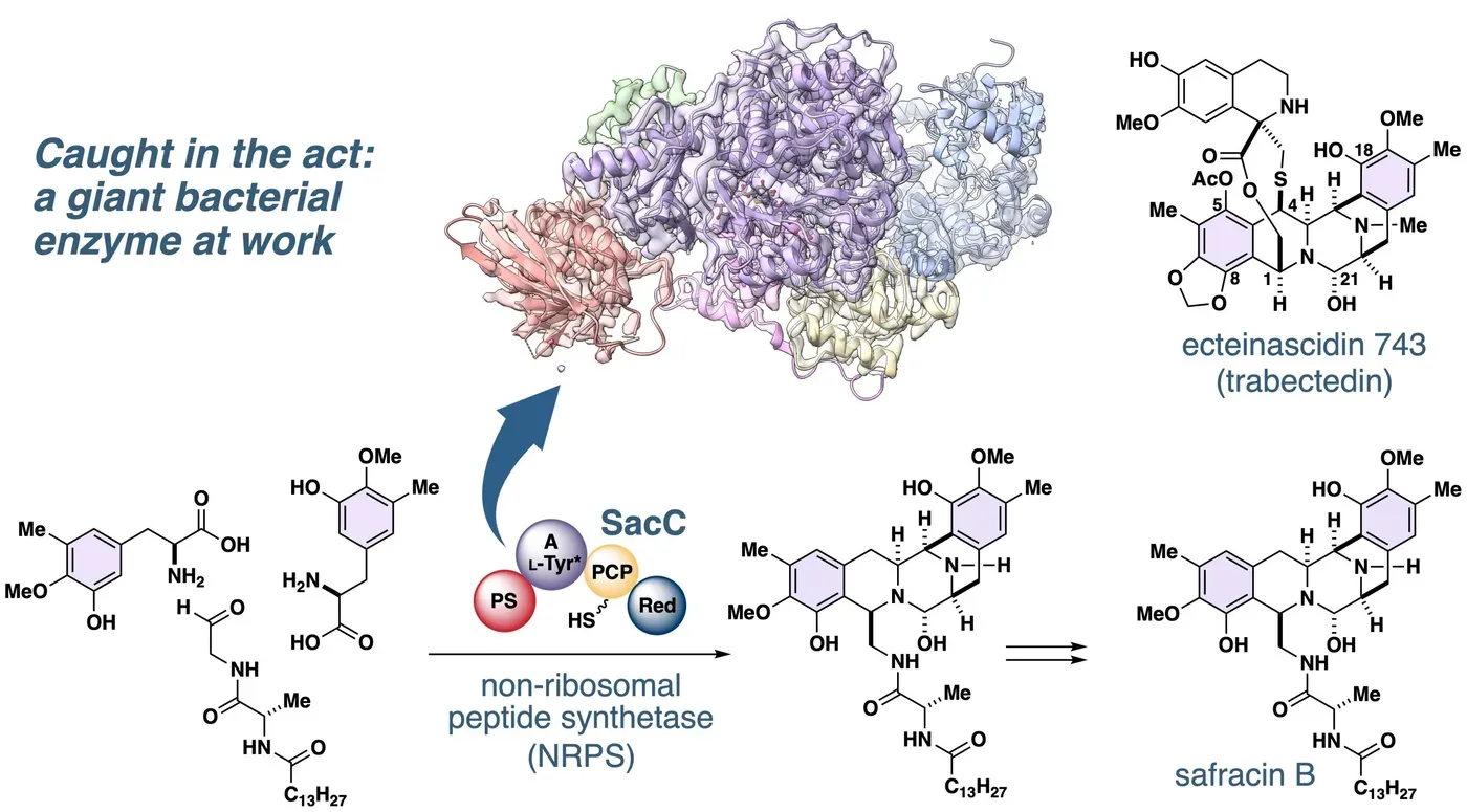
A team of researchers from Japan has made a groundbreaking discovery by visualizing an enzyme that plays a crucial role in building the complex core structure of safracin B, a key precursor to the cancer drug trabectedin.
The research team, led by Ryo Tanifuji and Hiroki Oguri from the University of Tokyo, and Yoshikazu Tanaka from Tohoku University, has successfully captured the image of SacC, an enzyme that assembles the five-ring core structure shared with trabectedin.
This achievement is significant not only because it provides a clear picture of the enzyme in action but also because the method used to visualize SacC may have far-reaching implications for research into related enzymes that produce valuable compounds.
The approach pioneered during this project has the potential to be applied to a wide range of enzymes, opening up new avenues for optimizing existing compounds and creating new ones.
Trabectedin is currently used to treat soft tissue sarcoma, and its production line begins with the bacterial compound safracin B, which SacC assembles within bacteria through a seven-step catalytic cascade.
This process involves stitching together two new carbon-to-carbon bonds in a single pass, producing the complex five-ring core scaffold that is also present in trabectedin.
The researchers tackled this complex problem by employing an interdisciplinary approach that combined chemistry and structural biology expertise.
To accurately visualize the process, they needed small molecules that mimicked the intermediate products formed during the enzyme's cascade reaction. By designing and synthesizing these molecules, the team could study the enzyme's behavior in a more controlled environment.
However, even with these synthetic intermediates, the researchers faced another hurdle: stabilizing the enzyme itself. This was crucial because the enzyme had a tendency to clump together in solution, making it difficult to obtain clear images.
The breakthrough came when the scientists discovered that a small partner protein called SacE significantly improved the enzyme's behavior in solution. This protein, encoded within the same bacterial genome as the enzyme, helped to disperse the enzyme molecules evenly.
With both challenges addressed, the researchers were able to flash-freeze their samples and use a cryo-electron microscope at Tohoku University to capture high-resolution images of the enzyme. The resulting three-dimensional model revealed intricate molecular details at a near-atomic level.
The researchers have managed to capture a crucial moment in the process of building a cancer drug scaffold using cryo-electron microscopy (cryo-EM). This early stage involves loading a building block onto an enzyme, which is then used to construct the final product.
However, the team's current model only shows the initial stages of this process and not the subsequent steps where the rings are closed and the finished product is released. They believe that capturing these later states will require more advanced probes and new methods for sorting images.
The scientists hope to refine their understanding of the enzyme cycle by obtaining a complete picture of how the cancer drug scaffold is built, which could ultimately lead to improved drug design and development processes.
Facts based on reporting originally published by Phys.org.
You may republish this story, in full or in part, if you credit News Central Site and link to it (licence CC BY 4.0). Photos are not included.