Brain Anatomy Mysteries Explored by Scientists
Neuroscientists Dr. Martin Wessendorf and Daniel Wang have shed new light on how the brain controls bodily functions.
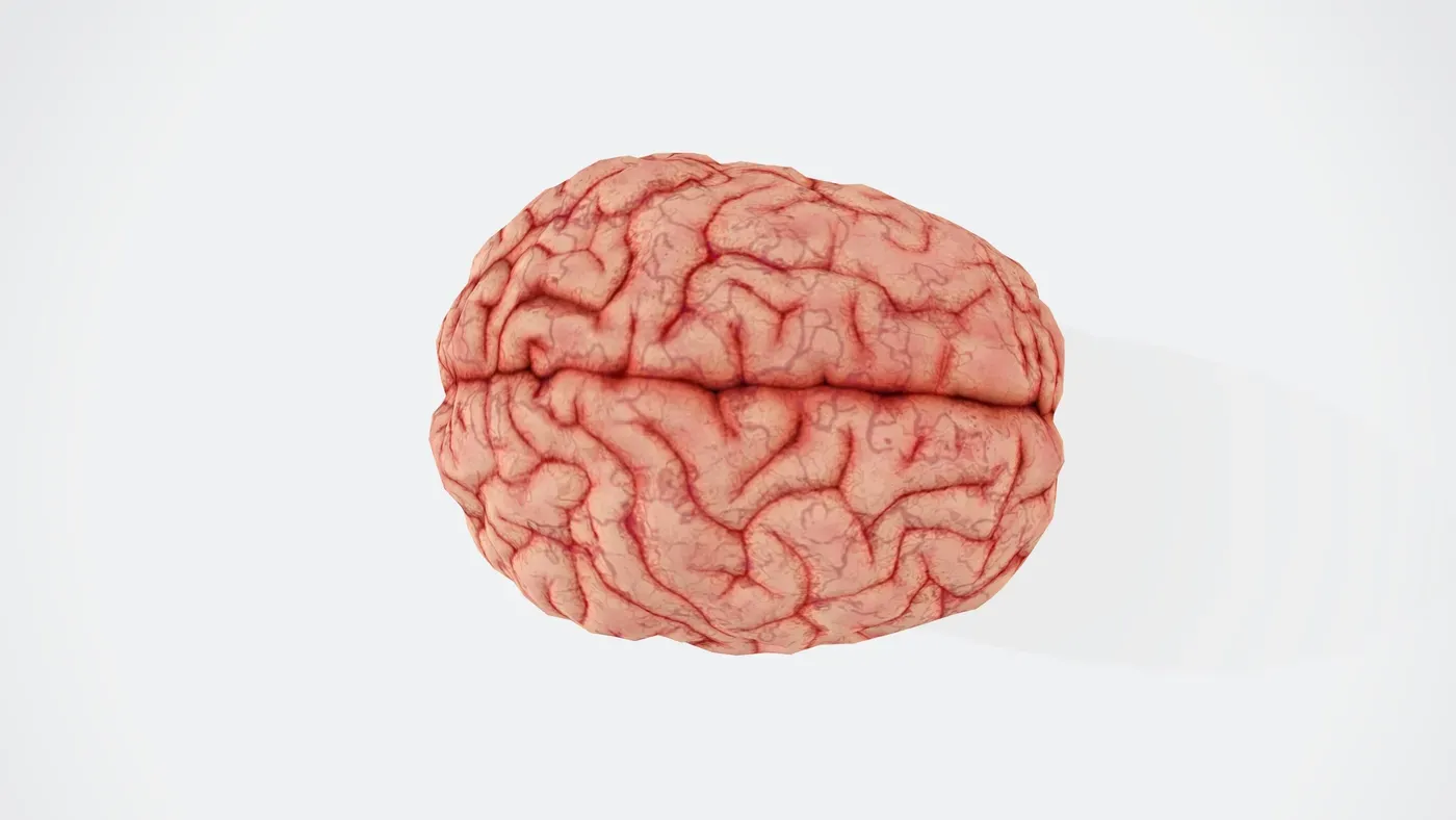
Neuroscientists offer fresh insights into a long-standing mystery of brain anatomy. Dr. Martin Wessendorf and Daniel Wang have been studying how the brain controls bodily functions, particularly in four-limbed animals. Their research focuses on cross-wired pathways, where one side of the brain connects with the opposite side of the body.
The team's latest work explores several key brain circuits that are involved in controlling movement and sensation. These pathways play a crucial role in our ability to interact with the world around us. In a new article published in Frontiers in Neuroanatomy, Wessendorf and Wang propose a novel hypothesis about how these cross-wired pathways might have evolved.
The story of brain anatomy begins with sensory input from our eyes. When light enters an eye, it forms an image on the retina that is reversed, meaning what we see to the right forms an image on the left side of the retina. This inverted image is then carried by the optic nerve to the optic chiasm, a crucial juncture in the brain where nerve fibers are sorted.
At the optic chiasm, the nerve fibers responsible for processing visual information from one half of the visual field are directed to the opposite half of the brain. This process is mirrored on the other side of the brain, with the right half of the brain receiving input from the left visual field. As a result, our visual inputs target an ancient structure called the optic tectum.
The team's hypothesis proposes that these cross-wired pathways may have evolved to facilitate more efficient communication between different parts of the brain. By connecting one side of the brain with the opposite side of the body, these pathways could enable faster and more coordinated responses to sensory stimuli.
Further research is needed to fully understand the evolution and function of these cross-wired pathways. However, Wessendorf and Wang's work provides valuable insights into the intricate mechanisms that govern our brain's control over bodily functions.
The midbrain contains a crucial structure known as the optic tectum, which is responsible for generating rapid responses to visual stimuli. This structure is present in all vertebrate species and appears to be one of the oldest neuroanatomical features in these animals. The optic tectum plays a vital role in processing visual information from each field of vision, mapping it to the opposite side of the brain.
Two neural pathways are responsible for transmitting physical sensations from most parts of the body. Both of these pathways cross the midline of the central nervous system and are found in all tetrapod species, which include mammals, birds, reptiles, and amphibians. This widespread presence suggests that these pathways have been conserved across evolution.
One of these pathways is known as the dorsal column pathway, which transmits sensations such as light touch, vibration, and body position from the limbs and body to the brain. The name "dorsal column" refers to its location in the spinal cord, where it ascends before connecting with another neuron that crosses the midline in the brainstem.
This pathway is not exclusive to tetrapods, having been observed in various species including amphibians, birds, mammals, and reptiles. Its presence across these groups highlights the importance of this pathway in transmitting physical sensations from the body to the brain. In contrast, fishes do not possess this pathway, suggesting that it may have evolved later in vertebrate evolution.
Another key pathway is the spinothalamic tract, which conveys information about pain, itch, and certain types of touch from the body to the brain. Unlike the dorsal column pathway, this tract crosses the midline at a different location, within the spinal cord itself. Once it has crossed, it ascends and transmits its sensory information to the thalamus.
The spinothalamic tract is found in various species including birds, mammals, reptiles, and even some cartilaginous fishes such as sharks and rays. Its contralateral organization means that it maps sensations from one side of the body to the opposite side of the brain, highlighting its importance in processing sensory information.
The brain's complex network of pathways plays a crucial role in controlling our movements and responses to stimuli. One such pathway is the tectospinal pathway, which enables us to react quickly to visual cues. For instance, when we're about to enter a crosswalk and catch a glimpse of an oncoming car from the corner of our eye, we instinctively turn towards it.
This reflex action is made possible by the way sensory inputs are mapped in the brain, with visual information being registered in the contralateral tectum. The optic tectum receives not only visual but also auditory and somatosensory inputs, which are then processed together to trigger a response. This pathway's ability to mediate responses to multiple types of stimuli highlights its importance in our daily lives.
The motor component of the tectospinal pathway originates from the deepest layers of the optic tectum and projects axons to the contralateral spinal cord. This connection enables the brain to send signals to the muscles, facilitating movement and reflexes. The presence of this pathway is not limited to mammals alone; it has been found in most tetrapods and some cartilaginous fishes.
Another pathway that facilitates limb control in animals is the rubrospinal pathway. Found primarily in mammals, this pathway originates from the red nucleus of the midbrain before projecting to the contralateral spinal cord. Its existence has also been reported in tetrapods and some cartilaginous fishes, indicating its significance across different species.
The presence of these pathways underscores the brain's remarkable ability to process and integrate various types of sensory information. By mapping sensations from one side of the body to the opposite side of the brain, these pathways enable us to respond quickly and effectively to our surroundings.
The corticospinal pathway in humans is a unique feature that enables fine motor control. This pathway diverges from the general vertebrate pattern, where the telencephalon, the front part of the brain, contains a six-layered cerebral cortex responsible for higher-order functions.
In mammals, including primates and humans, this pathway can modulate either somatosensory input or motor output. The corticospinal tract travels from the cerebral cortex to the spinal cord, where it influences sensations entering through the body or controls muscle movements.
The function of the corticospinal pathway varies across species, with some mammals using it primarily for somatosensation and others, like primates, utilizing it for fine motor control. This distinction highlights the adaptability of this neural circuit over evolutionary time.
The brain's organization is thought to have been influenced by ancient visual pathways that bias central nervous systems toward contralateral organization in tetrapods. The presence of an optic chiasm and a contralaterally organized optic tectum suggests that early jawless fishes had a similar brain structure.
These findings imply that the corticospinal pathway's role in fine motor control may have evolved from an earlier function related to somatosensation. This idea can be tested by examining fossilized remains of ancient fish skulls, which may provide evidence for the presence of both an optic chiasm and an optic tectum.
Further research is needed to confirm this hypothesis, but it offers a new perspective on the evolution of fine motor control in humans and other mammals.
The right side of the brain controls the left side of the body due to a fundamental principle of evolution: it often builds upon existing structures rather than inventing new ones. This means that the organization of the visual system, particularly in the optic tectum, may have laid the groundwork for the sensory and motor pathways that followed. As a result, contralateral organization - where one side of the brain controls the opposite side of the body - may not be an adaptation per se, but rather a byproduct of evolution working with what it already had.
This idea challenges the conventional understanding that contralateral organization evolved as a specific adaptation to improve motor control. Instead, it suggests that this trait emerged because evolution was constrained by its existing infrastructure and materials. In other words, the brain's organizational principles were shaped by its pre-existing visual system, which in turn influenced the development of sensory and motor pathways.
The optic tectum is a key structure in the visual system that processes visual information from each eye. Its organization may have set the stage for the contralateral control observed in other parts of the brain. This perspective offers a new way of thinking about the evolution of fine motor control, not just in humans but also in other mammals.
This hypothesis highlights the intricate and interconnected nature of brain development and evolution, where seemingly unrelated systems can influence one another in complex ways.
Facts based on reporting originally published by Phys.org.
You may republish this story, in full or in part, if you credit News Central Site and link to it (licence CC BY 4.0). Photos are not included.