A sunlight-powered breakthrough
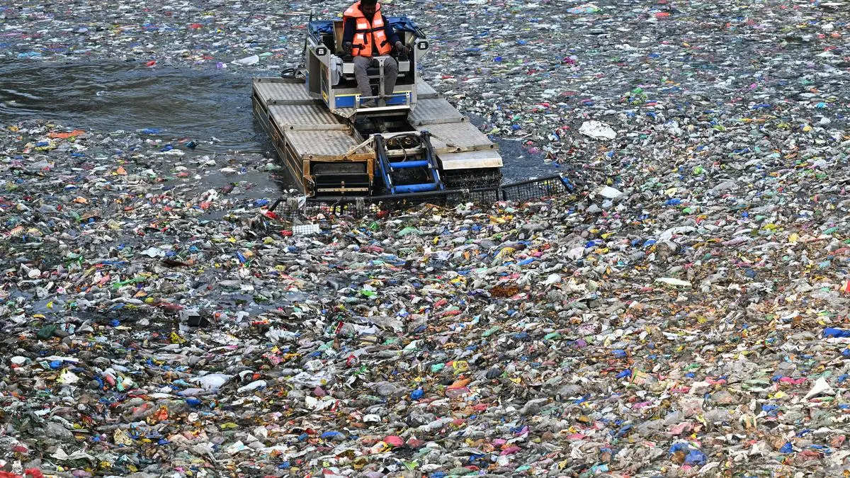
Plastic waste, notably microplastics, has been discovered throughout most of the planet’s ecosystems, elevating issues about threats to terrestrial and marine life and to human well being as effectively. To sort out this downside, researchers on the University of Waterloo, Ontario, Canada, developed a means, utilizing daylight, to flip plastic waste into acetic acid, the primary ingredient of vinegar.
White rot fungus (Phanerochaete chrysosporium) is understood for its skill to break down lignin, one of many hardest polymers present in wooden. It makes use of enzymes that generate extremely reactive chemical species able to dismantling complicated carbon buildings. Yimin Wu and his colleagues on the college puzzled if a artificial materials may mimic this technique. The staff developed a bio-inspired cascade photocatalysis utilizing iron-doped carbon nitride because the catalyst. Carbon nitride is a semiconductor that absorbs seen mild.
“Rather than forming nanoparticles, each iron atom is isolated and embedded within the carbon nitride structure. This atomic precision is crucial. Each iron atom behaves like an active site in a natural enzyme, maximizing efficiency while maintaining stability,” Wu wrote in The Conversation, a journal, describing the work.
The system works via a cascade of light-driven reactions, he defined. Under daylight, and within the presence of hydrogen peroxide, the iron websites activate the peroxide to generate hydroxyl radicals. A radical is an atom, molecule, or ion that has at the very least one unpaired electron, which makes it extremely chemically reactive.
These radicals assault the lengthy carbon chains that make up plastics, like polyethylene (utilized in plastic luggage), polypropylene (meals containers), PET (drink bottles), and even PVC (pipes and packaging). The polymers are progressively oxidised and damaged down into smaller molecules, finally forming carbon dioxide (CO2). Rather than permitting this CO2 to escape, the identical catalyst then makes use of daylight to scale back the CO2 into acetic acid.
In different phrases, the carbon in plastic waste is first oxidised after which reassembled into a new, beneficial molecule. “Essentially, this approach breaks down plastic and converts the resulting carbon into a commodity chemical in a single system. This distinguishes it from most existing recycling technologies,” Wu mentioned.
Since the response takes place in water, it makes the method significantly related for plastic air pollution in aquatic environments. The researchers additionally examined the method with actual world waste streams containing polluted plastic and mixtures of various sorts. Significantly, in contrast with many chemical recycling strategies that require heating plastic to a number of hundred levels Celsius, this response proceeds at room temperature and regular atmospheric strain.
Acetic acid is broadly utilized in meals manufacturing, chemical manufacturing, and vitality purposes. Currently, most acetic acid is produced via an energy-intensive course of course of referred to as methanol carbonylation, whereby methanol is reacted with carbon monoxide at excessive temperatures. “By mimicking the way enzymes control reactivity at precise metal centres, we can achieve complex chemical transformations under mild conditions using sunlight as the energy source,” Wu said.
While this method remains to be on the laboratory stage, the staff envisions that it might be tailored for scalable, solar-driven recycling and environmental clean-up and the photocatalytic upcycling system will be additional enhanced via strategic supplies engineering and manufacturing processes.

A affected person present process CT scan. A examine by the International Atomic Energy Agency discovered vital variation within the radiation dosage obtained by sufferers from diagnostic exams for coronary artery illness.
| Photo Credit:
Okay.R. Deepak
IAEA flags worldwide variation in radiation doses from diagnostic imaging
A examine by the International Atomic Energy Agency (IAEA) has discovered vital variation within the radiation dosage obtained by sufferers from diagnostic exams for coronary artery illness. The examine, which was funded and performed underneath the IAEA-coordinated analysis venture IAEA Noninvasive Cardiology Protocols Study (INCAPS4) was carried out in affiliation with Columbia University, New York City.
The findings underscore an pressing want for improved coaching, standardised protocols, and up to date gear, significantly in low- and middle-income nations, the place the information counsel that radiation doses might be lowered with out compromising check outcomes. The outcomes have been printed not too long ago in The Journal of the American Medical Association.
The examine analysed information from greater than 19,000 sufferers at 742 centres in 101 nations, gathered over a 9-week interval in 2023, making it the biggest and most complete world evaluation of radiation publicity from non-invasive cardiac imaging.
Researchers examined radiation doses from broadly used imaging strategies, together with nuclear cardiology exams and computed tomography (CT) scans of the center. While many centres may maintain radiation publicity inside advisable limits, the examine discovered marked variations between nations, areas, and earnings ranges, with some folks receiving greater doses than others for a similar check.
The examine discovered that median radiation doses various broadly by imaging modality, earnings ranges, and geography. Optimised protocols and use of newer know-how, which regularly ship clearer pictures, have been persistently related to decrease affected person publicity. Patients in low- and middle-income nations usually obtained considerably greater doses, significantly for coronary CT angiography, a check that’s more and more used due to CT’s availability and technological enhancements.
“These differences are not inevitable,” mentioned Andrew J. Einstein, the examine’s principal investigator and corresponding creator. “In many cases, the technology and knowledge to reduce dose already exist. The challenge is ensuring that they are applied consistently and equitably across the world.” The authors emphasised that lowering radiation doses doesn’t imply lowering diagnostic high quality.
“This research provides critical evidence that can inform national policies and international action,” mentioned Diana Paez of the IAEA, the senior investigator and co-lead of the examine. The examine factors to the significance of peer-to-peer information sharing, improvement of regional and world dose reference ranges, and nearer collaboration between regulators, skilled societies, and trade, an IAEA launch mentioned.
. He won the Nobel Prize in Chemistry Prize in 2014 along with Eric Betzig and Stefan Hell “for the development of super-resolved fluorescence microscopy”.")
W.E. Moerner (an undated {photograph}). He gained the Nobel Prize in Chemistry Prize in 2014 together with Eric Betzig and Stefan Hell “for the development of super-resolved fluorescence microscopy”.
| Photo Credit:
Linda A. Cicero/Stanford News/Handout by way of Reuters
Label-free microscope for nanoscale view inside dwelling cells
By combining two microscopy strategies, researchers at Stanford University have developed a one-of-a-kind instrument that may reveal cell buildings interacting in actual time at an unprecedented decision of 120 nanometres, the best achieved with out using fluorescent labels.
This new “label-free” know-how, referred to as Interferometric Image Scanning Microscopy (iISM), permits commentary of mobile buildings of their wider context, together with their responses to intrusions, reminiscent of pathogens or medication. The advance was printed within the journal Light: Science and Applications.
iISM’s capabilities open up new avenues in a vary of life science fields, from understanding illness mechanisms and creating medication to investigating relationships between crops and microbes.
Even although the decision is just not as positive as different forms of specialised microscopes, iISM permits the viewing of many buildings without delay for a lengthy period. In distinction, strategies that depend on fluorescence to label buildings sometimes can spotlight solely a few goal buildings at a time. Also, fluorescence can finally “bleach” or put on off. These labels will also be troublesome to introduce and, in some circumstances, might change the behaviour of the buildings they tag.
The distinction between earlier high-contrast label-free approaches and iISM is that it could possibly function at a considerably decrease illumination (lower than a microwatt), which reduces the chance of photodamage to dwelling cells and makes it much less seemingly to disturb the tiny, delicate buildings.
iISM manages to obtain greater decision and sensitivity by combining some great benefits of two completely different microscopy strategies which are the respective areas of experience of the 2 scientists who created the method and authored the paper on it. W.E. Moerner gained the 2014 Nobel Prize in Chemistry for his work on super-resolution fluorescence microscopy and Michelle Kueppers’ doctoral work focussed on “interferometric scattering (iSCAT) microscopy”.
In iSCAT microscopy, a laser shines on a cell, and the tiny buildings inside scatter a number of the mild. A second laser beam is then used to amplify the weaker scattered mild and make it robust sufficient to be detected in order that the small buildings will be seen.
The key advance in iISM is the mix of the iSCAT technique with an tailored idea from next-generation confocal microscopes. While conventional confocal microscopes use a pinhole and a single detector to give attention to goal buildings, superior variations of those microscopes use camera-based array detectors, which seize many views of the identical space.
iISM makes use of a sort of array detector that collects extra mild than the pinhole and single detector mannequin. This gives elevated depth and precision. It works comparable to the way in which two human eyes each soak up data to distinguish the foreground from background besides that iISM makes use of tens to lots of of views from an array detector. The researchers then developed a technique to mix these measurements into sharper, higher-contrast pictures.
iISM will, after all, not change using fluorescence microscopy, which has yielded insights into the life sciences for many years. “Every method has its advantages and disadvantages, and we believe in a complementary implementation in the future,” mentioned Kueppers. “If we use the strengths of fluorescence for molecular specificity and the strength of iISM for label-free context and dynamics, we can really start tackling questions that have been difficult to address before.”
Also Read | Plastic pollution is surging. What are governments doing?
Also Read | Atoms in industry