Brain-IT uses AI to recreate brain scan data into vivid images
A new AI tool called Brain-IT has been developed to transform brain scan data into accurate visual representations.
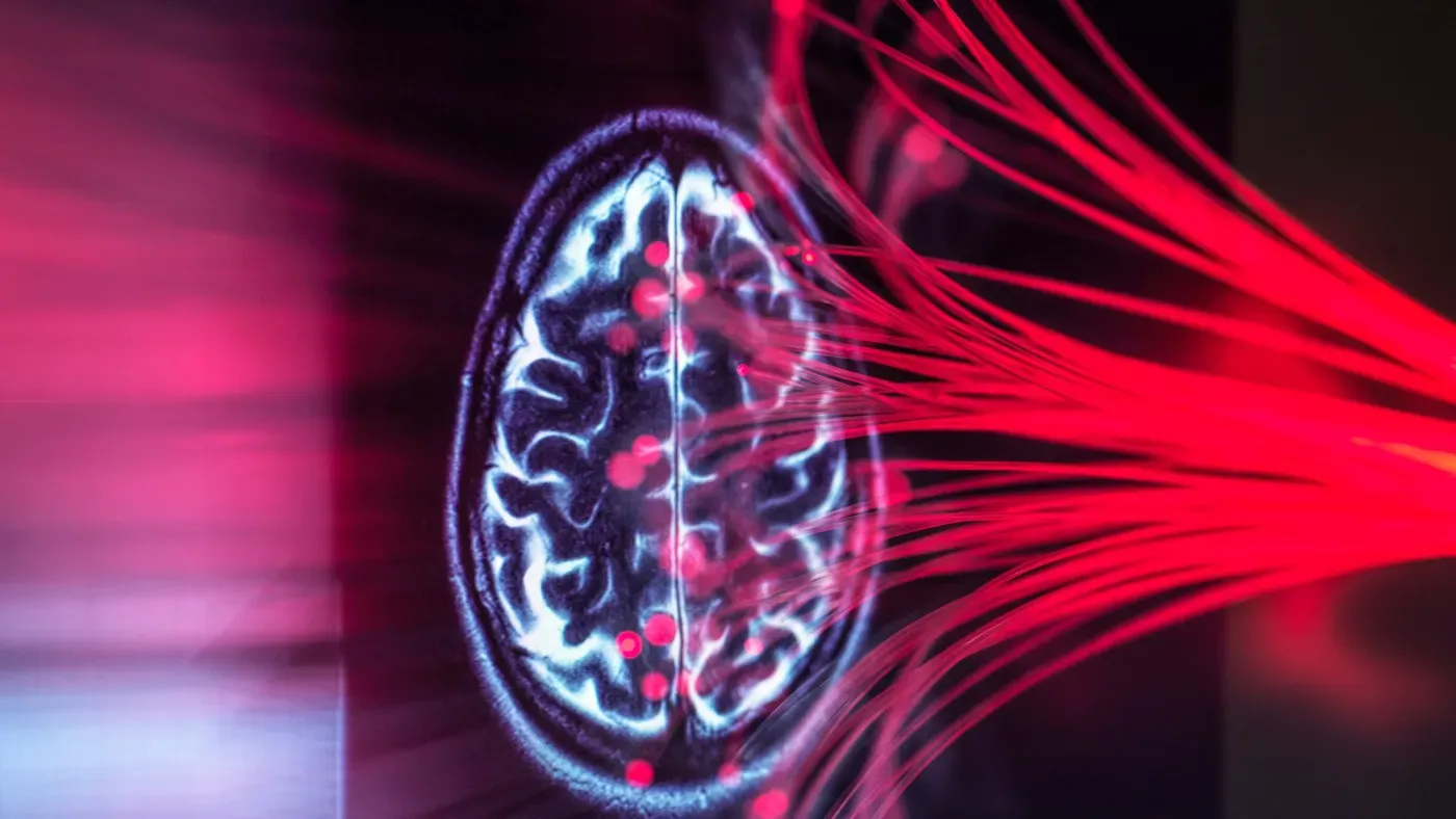
Brain-IT, a cutting-edge AI tool, has been developed to transform brain scan data into vivid images, displaying an uncanny ability to recreate visual stimuli with remarkable accuracy.
The system relies on functional magnetic resonance imaging (fMRI) scans to measure activity in the visual cortex, the brain region responsible for processing visual information. By analyzing these patterns of brain activity, Brain-IT can generate remarkably accurate depictions of the original images.
Researchers used a computer model to predict both the overall layout and high-level details of an image based on its content. This information is then fed into an AI-powered image generator, which produces the final visual representation of the original stimulus.
The technique builds upon an existing approach called the Universal Brain Encoder, which works in reverse by predicting fMRI responses from individual "voxels, tiny 3D units within the brain scan.
This process allows researchers to pinpoint exactly what each voxel is sensitive to in images, whether it's edges, textures or specific locations.
The Universal Brain Encoder has been designed to accommodate diverse datasets and participants, unlike traditional image-to-fMRI systems that are typically custom trained for each individual case.
This flexibility is due in part to the system's ability to learn from a wide range of data sources, allowing it to adapt and generalize across different MRI systems with varying magnetic-field strengths.
A key aspect of this process involves the identification of specific patterns within brain activity measurements, which can then be used to inform the reconstruction of visual images.
The Universal Brain Encoder achieves this by dividing thousands of tiny brain-scan measurements from the visual cortex into 128 distinct groups based on their typical associations with different types of visual information.
These groups are allowed to share and combine their insights, enabling the system to synthesize a more comprehensive understanding of an image's content.
The other type of information captured by this AI is structural in nature, focusing on the overall visual layout and arrangement of forms within an image.
This can include aspects such as the outline of a specific object or its position within the frame, providing a foundation for the generator to build upon.
In contrast to beginning with pure noise, the structural prediction serves as a starting point for the image generation process, while the semantic information guides the removal of static until the final image emerges.
The AI's ability to recreate visual content is not without limitations, however, and it cannot reproduce memories or imagined scenes from brain activity data.
We're not reading people's thoughts, Michal Irani emphasized, underscoring that the tool merely interprets visual brain activity.
A high-scoring or realistic-looking output from this AI system should not be mistaken for an exact replica of what someone saw.
The final stage of processing involves feeding the visual brain activity into an off-the-shelf diffusion generator trained on a vast library of images to produce a generated picture.
This type of model can create plausible images that not only generate high-quality textures but also sharpen facial features and fill in missing details, making them more polished and convincing.
The system's beautification" introduces details that aren't necessarily derived from the brain data itself, relying on patterns learned from photographs to add realism to the generated image.
However, this process is limited to reconstructing seen images, as it relies on the brain's processing of visual information rather than memories or imagined scenarios.
The brain's visual activity patterns, though vague and influenced by networks involved in memory and thought, hold some clues about how visual information is processed in different regions.
To unlock this potential, researchers rely on fMRI scanning, which, although expensive and cumbersome, provides the necessary data for accurate results. However, a more accessible alternative is being explored: electroencephalogram (EEG) technology, which uses electrodes to collect data from the head, but its development is still in its early stages.
This innovative system could revolutionize brain research by mapping visual information across different regions and revealing how individuals' patterns differ. Ultimately, such tools may shed light on neurological, neurodevelopmental, and psychiatric conditions, paving the way for new medical applications that are currently being pursued.
Facts based on reporting originally published by Live Science.
You may republish this story, in full or in part, if you credit News Central Site and link to it (licence CC BY 4.0). Photos are not included.