
The analysis is opening new frontiers in pest management and evolutionary biology.
An worldwide group of scientists has uncovered a unusual tubular construction inside Profftella, a bacterium that lives in shut partnership with a main citrus pest. Nothing like this construction has ever been documented in any residing organism. The discovery, made potential via superior microscopy, may open the door to new methods for pest management and supply contemporary insights into the evolution of life.
The analysis was carried out by scientists from Pusan National University, the National Institute for Physiological Sciences, Kobe University, and Toyohashi University of Technology. They recognized the bizarre construction inside Candidatus Profftella armatura, a symbiotic bacterium that inhabits the Asian citrus psyllid (Diaphorina citri). Their findings have been revealed in NPJ Imaging on September 18, 2025.
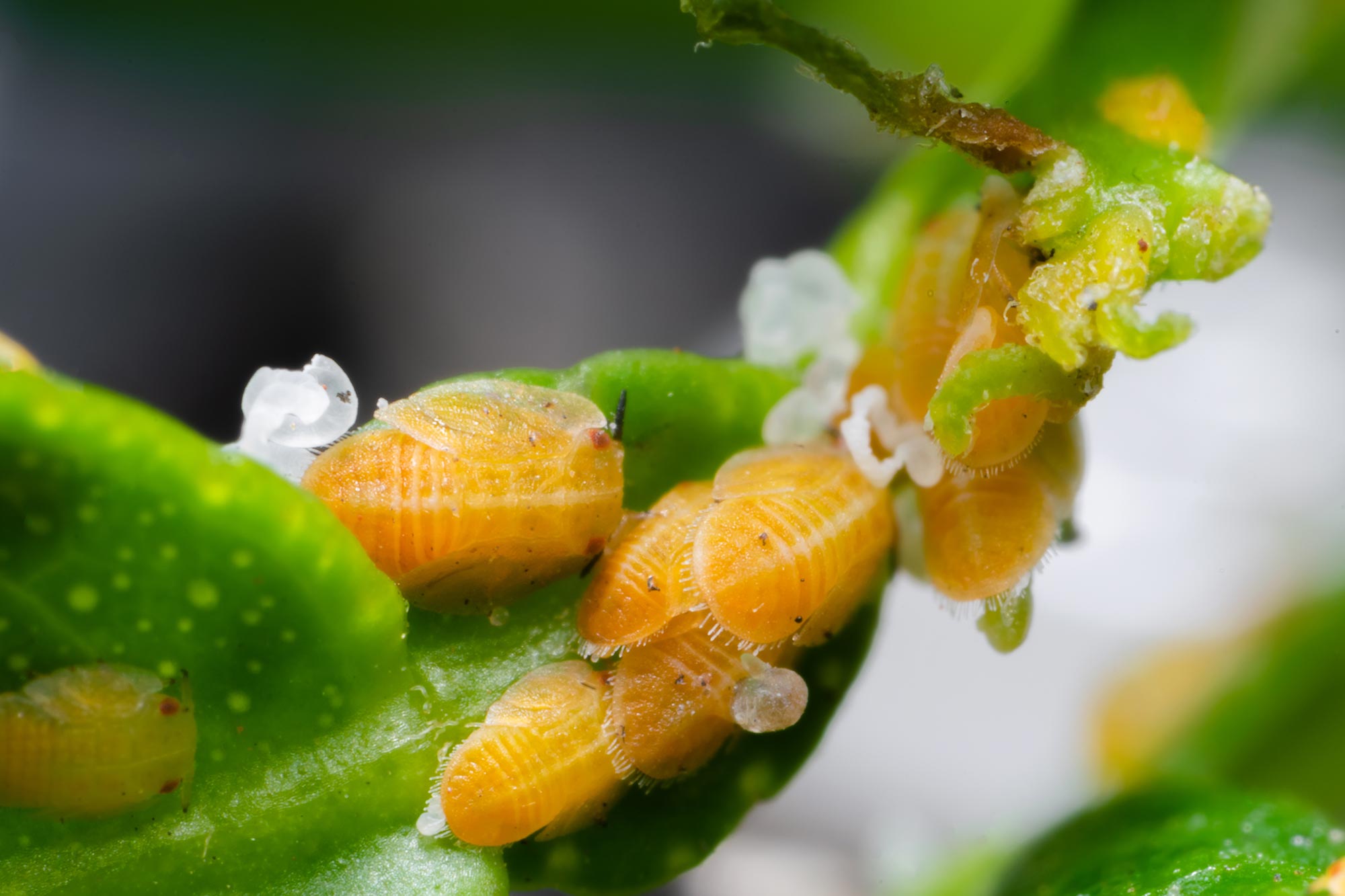
The Asian citrus psyllid is a damaging insect that inflicts main harm on citrus crops across the globe, decreasing harvests and driving up costs. This pest carries Profftella, a bacterium handed down via generations of psyllids. The microbe performs a essential position in the insect’s survival and produces poisonous substances that assist protect its host from pure enemies.
Discovery of an Unprecedented Tubular Structure
Using superior 3D electron microscopy, the workforce revealed that Profftella cells, that are themselves extraordinarily elongated, measuring as much as over 100 micrometers in size, include a number of elongated tubular constructions every measuring tens of micrometers in size. It was revealed that these tubes have a diameter of roughly 230 nm and are composed of 5 to six right-handed helical fibers forming a twisted construction. They have been additionally discovered to occupy a constant proportion of the Profftella cell quantity.
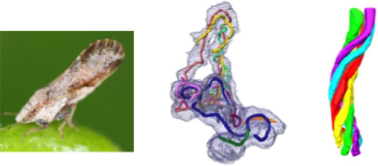
“Typically, bacteria lack such complex organelles. Moreover, I was surprised to find that this tube is so stable and robust that it maintains its shape during high-vacuum electron microscopy observation without chemical fixation or embedding,” explains Assistant Professor Chihong Song from Pusan National University, the article’s first writer.
Further analyses utilizing varied strategies, together with optical microscopy, revealed that these tubes include quite a few ribosomes, the mobile equipment liable for protein synthesis.
“Based on this, the tubes may be involved in protein synthesis. Given their sturdiness, they could also provide physical support to the elongated Profftella cells, and maybe even act like a scaffold for material transport—kind of like the cytoskeleton in eukaryotic cells,” says Associate Professor Atsushi Nakabachi from Toyohashi University of Technology, a corresponding writer of the research.
Rethinking Bacterial Complexity
The analysis workforce emphasizes that this construction represents a particularly uncommon instance of an organelle discovered in micro organism.
This vital discovery challenges the traditional view of micro organism as easy life types and affords new insights into the evolution of mobile constructions. At the identical time, it opens promising avenues for growing selective pest management methods concentrating on the Asian citrus psyllid, with necessary implications for agriculture.
Reference: “Enigmatic tubular ultrastructure in the bacterial defensive symbiont of the Asian citrus psyllid Diaphorina citri” by Chihong Song, Junnosuke Maruyama, Kazuyoshi Murata, Toshinobu Suzaki and Atsushi Nakabachi, 18 September 2025, npj Imaging.
DOI: 10.1038/s44303-025-00107-w
This work was supported by the Japan Society for the Promotion of Science (https://www.jsps.go.jp) KAKENHI (grant numbers 21687020, 26292174, 20H02998 and 25K02023 to AN), the Collaborative Study by High Voltage Electron Microscopy Program (2015-502, 2016-502) of National Institute for Physiological Sciences to AN, and a National Research Foundation of Korea (NRF) grant RS-2024-00440289 to CS.
Never miss a breakthrough: Join the SciTechDaily newsletter.